
Divisions Euglenophyta, Phaeophyta, Chrysophyta Plantlet
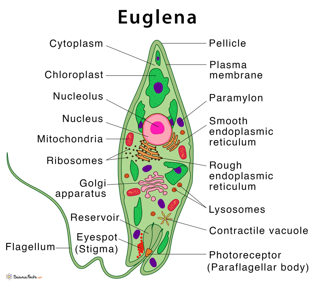
Description. Euglena Anatomy Diagram.svg. English: The anatomy of the protist Euglena Euglena are unicellular, flagellate protists of the genus Euglena and kingdom Eukarya. Able to photosynthesize with their chloroplasts and also capable of accessing food from outside sources, they are both autotrophic and heterotrophic.

How To Draw Euglena step by step tutorial YouTube
Euglena Euglena are one-celled protozoans with a whip-like flagellum that they use to propel themselves through their aquatic environment. Some euglena species have chlorophyll, so can produce their own food through photosynthesis. ©Sheri Amsel www.exploringnature.org nucleus flagellum plasma membrane golgi apparatus rough endoplasmic.

Euglena Diagrams 101 Diagrams
Structure. Euglena is a unicellular organism with a complex internal structure that includes a contractile vacuole that can expel water and a red 'eyespot' . Photosynthetic forms contain a chloroplast. They possess two flagellae, one long, one short, which can allow the organisms to move. Euglena are also able to move by means of changing its.
Euglena Definition, Structure, & Characteristics with Diagram
On the right is a diagram of a Euglena displaying its Organelles, which include: Flagellum- A long, mobile filament that the Euglena uses to propel itself in its environment Reservoir- The part used for storage of nutrients
Euglena under a microscope anatomy, reproduction & facts Rs' Science
Euglena Structure with Diagram and Characteristics Morphology and Anatomy Euglena has an elongated cell measuring 15-500 micrometres Mostly green in colour due to the presence of chlorophyll pigment Some of the species of euglena contain carotenoid pigments, which give it distinct colour like red Euglena is unicellular having one nucleus
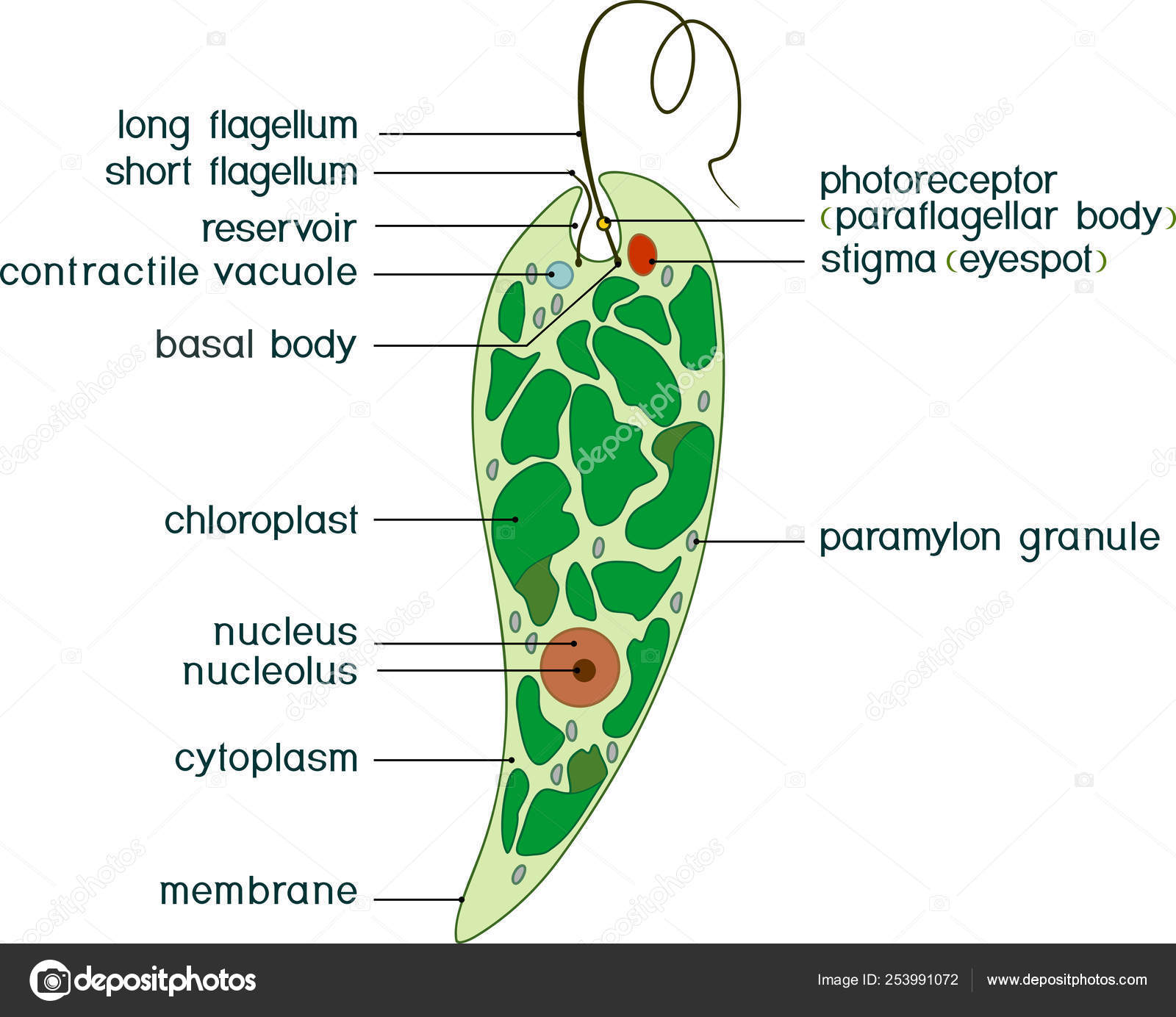
Diagram Euglena Structure Euglena Viridis Titles Stock Vector Image by ©mariaflaya 253991072
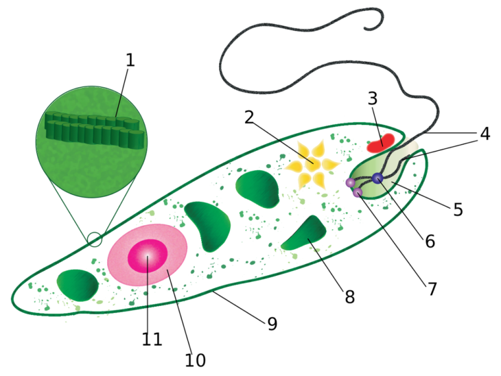
The Euglena Diagram is typically presented as a labeled image, with each structure illustrated with a different color or shape. Students can then use these labels to create flashcards, diagrams, or graphic organizers to review the Euglena's structure and functions. Additionally, teachers can ask students to perform hands-on activities, such.

Well Labelled Diagram Of Euglena Euglena Under A Microscope Anatomy Reproduction Facts Rs
Structure Euglena is an elongated or spindle-shaped cell with a size around 15-500 x 10 -6 m. Parts Euglena The internal structures found in a typical photosynthetic Euglena are as follows: Pellicle: A thin, flexible membrane that supports the plasma membrane and helps them to change shape

Euglena Structure And Function Euglena 3D model by Visartech (visartech) [d92e880
The following 15 files are in this category, out of 15 total. Augentierchen 440x600.jpg 440 × 599; 47 KB Euglena - schema.svg 400 × 600; 51 KB Euglena Anatomy Diagram.svg 600 × 450; 6.06 MB Euglena diagram gl.jpg 800 × 524; 104 KB Euglena diagram.jpg 800 × 524; 150 KB Euglena gracilis.jpg 500 × 500; 147 KB Euglena gracilis.svg 202 × 635; 216 KB

Euglena Diagrams 101 Diagrams
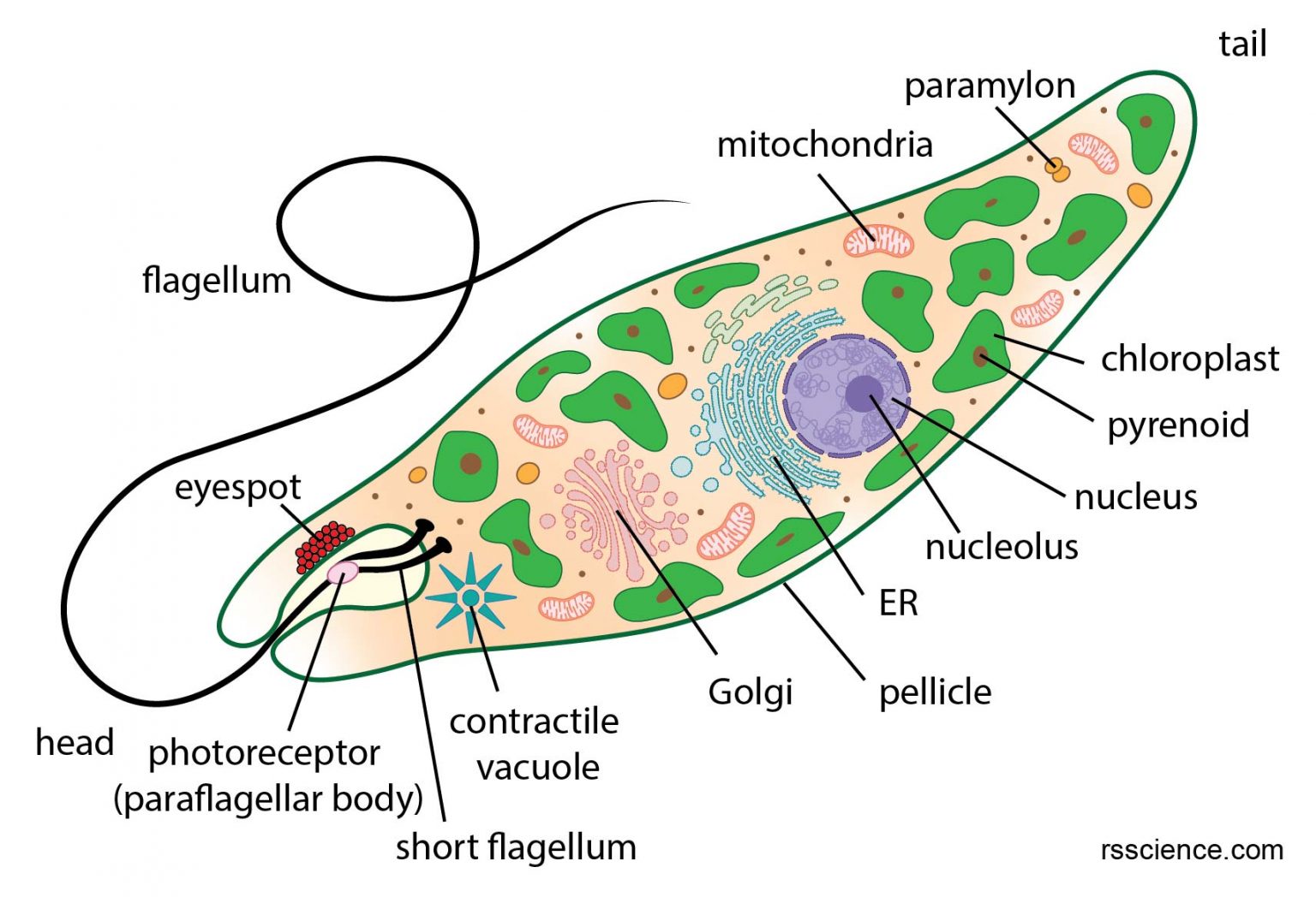
Euglena (Greek: eu = true, glene = eye-ball) is a genus of single cell eukaryotes with flagella, and they can be found in freshwater pond and ditches. Euglena gracillis is one of the species that has been used as a model organism for studying cell biology in the lab.

Euglena Diagrams 101 Diagrams
The Diagram and Characteristics of Euglena The Morphology and Anatomy of Euglena Euglena has a unique, elongated cell structure that measures between 15-500 micrometres. The organism is primarily green due to the presence of chlorophyll pigment. Some Euglena species contain carotenoid pigments, which give them a distinct color, such as red.
/euglena-57ee66383df78c690faf9a1b.jpg)
Anatomy and Reproduction of Euglena Cells
1. A small free living and freshwater form. ADVERTISEMENTS: 2. Body spindle shaped and usually green in colour and is surrounded with tough & elastic membrane - the pellicle. 3. In the middle of anterior end lies a cytostome leading into a small cytopharynx which dialates behind into a reservoir. 4.

anatomy of euglena
Euglena are tiny protist organisms that are classified in the Eukaryota Domain and the genus Euglena. These single-celled eukaryotes have characteristics of both plant and animal cells. Like plant cells, some species are photoautotrophs (photo-, -auto, -troph) and have the ability to use light to produce nutrients through photosynthesis.

Anatomy Of Euglena Stock Illustration Download Image Now iStock
Euglena are characterized by an elongated cell (15-500 micrometres [1 micrometre = 10 −6 metre], or 0.0006-0.02 inch) with one nucleus, numerous chloroplasts (cell organelles that contain chlorophyll and are the site of photosynthesis), a contractile vacuole (organelle that regulates the cytoplasm), an eyespot, and one or two flagella.

Structure of a euglena Royalty Free Vector Image
Structure, Morphology and Classification Euglena are single celled organisms that belong to the genus protist. As such, they are not plants, animal or fungi. In particular, they share some characteristics of both plants and animals.
.PNG)
Euglena Structure And Function Euglena 3D model by Visartech (visartech) [d92e880
January 25, 2022 by Jamar Williams Table of Contents [ show] What is Euglena? Euglena is a genus classification of organisms that exhibit characteristics of both plants and animals. The euglena movement and structure are unique. These organisms' movement is based on the structural possession of a flagellum and pellicle.
Euglena Diagrams Labeled 101 Diagrams
No higher resolution available. Euglena_diagram.jpg (800 × 524 pixels, file size: 150 KB, MIME type: image/jpeg) File information. Structured data. Captions. Captions. English. Add a one-line explanation of what this file represents. Summary[edit]